Conditions & Treatments

Pain Conditions

Click on each condition to open


Arthritis is a chronic condition that affects your joints. There are over 100 types of arthritis, but many people struggle with the symptoms of two specific types — rheumatoid arthritis and osteoarthritis.

Rheumatoid arthritis is an autoimmune disorder that causes your body to mistakenly attack the healthy lining of the joints. This results in inflammation and irritation that can become painful and debilitating.

In osteoarthritis, the cartilage that protects the ends of the bones deteriorates, causing the bones that form a joint to rub together. This friction can cause pain and stiffness that makes it difficult to move the affected joint.

For advanced arthritis that causes chronic pain, your provider may suggest joint injections. This treatment delivers anti-inflammatory medications and a local anesthetic into the joint to reduce inflammation and disrupt the pain signals.

It’s estimated that as many as 80% of adult Americans experience back pain during their lifetime. Back pain can develop due to injuries, trauma, or degenerative diseases that progress as you get older.

Some of the most common causes of persistent back pain include:

  • Sciatica
  • Scoliosis
  • Herniated or bulging discs
  • Osteoarthritis
  • Facet joint syndrome
  • Osteoporosis

If back pain is severe enough to interfere with your life, you may benefit from steroid injections to reduce the inflammation that’s irritating your nerves. If severe pain is the result of a herniated disc or other spinal condition, epidural steroid injections may be helpful. These injections deliver anesthetic and steroid medications directly into the epidural space of the spine to relieve pain and decrease inflammation.


CRPS is a chronic pain disorder that typically affects one limb. There are two types of CRPS. CRPS-I is caused by an injury like a sprain or fracture. CRPS-II develops due to nerve damage. Their characteristic symptom is that the initial pain persists long after the injury or nerve damage heals. Additionally, the chronic pain is disproportionate to the original injury.

Both types of CRPS share the same symptoms. The primary symptom is persistent pain that’s often severe. Your pain begins at the site of your initial injury, which is usually a hand, foot, finger, or toe, then it may spread to include the entire arm or leg.

The affected area may become hypersensitive, making even a slight, gentle touch feel painful. The nerve damage underlying both types of CRPS affects your blood vessels, resulting in changes in your skin color, texture, and temperature.

Most patients also experience:

  • Swelling in the affected limb
  • Joint stiffness
  • Abnormal sweating near the injury
  • Changes in nail and hair growth on the affected limb
  • You may gradually develop muscle symptoms like spasms, atrophy, or loss of coordination.

When your symptoms don’t respond to conventional treatment like medications, the doctors at the Pain Institute of Southern Arizona (PISA) may recommend treatments that stop the nerves from transmitting pain. These are only two examples of their specialized pain treatments:

Sympathetic nerve block
When any part of your body is injured, sensory nerves carry signals to the spinal cord, and from there to the brain. Clusters of sensory nerves from one part of your body enter your spinal cord at the same spot. Your doctor can inject a local anesthetic that blocks those nerve signals, stopping them from reaching your brain. As a result, you don’t feel the pain caused by CRPS. Your pain relief typically lasts for several hours after the anesthetic normally wears off, and with additional injections, you may get longer-lasting relief.

Spinal cord stimulator
A spinal cord stimulator is a medical device that uses a mild electrical signal to block or mask the nerve signals. You’ll have a trial period to be sure it works, then the generator and electrodes are implanted so they can stay in place and provide long-term pain relief.


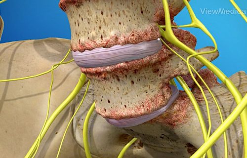
Like other soft tissues in your body, the discs between spinal vertebrae are susceptible to degenerative changes. Over the years, the discs dehydrate, thin out, and can’t continue to do their normal job of cushioning your vertebrae and absorbing shock. Daily wear-and-tear also damages the tough outer covering.

These changes eventually make the disc collapse. When that happens, the spine can become unstable. Additionally, the vertebrae above and below the disc move closer together, leading to the development of bone spurs.

Any time a disc collapses, vertebrae move out of their normal alignment or bone spurs develop, and the nearby nerves can become compressed. The damaged nerve causes pain that may be a constant ache or a severe, sharp pain.

The most common symptoms include:

  • Pain in the affected area and possibly radiating down an arm or leg
  • Pain that’s worse when sitting, bending, lifting, or twisting
  • Pain that improves when you lie down or change positions
  • Periods of pain that come and go
  • Tingling or numbness in your arm or leg
  • Weakness in arm or leg muscles
  • If you develop spinal instability, you may also develop painful muscle spasms.

Treatment for disc degeneration often begins with physical therapy customized to strengthen your spine, improve movement, and diminish pain and swelling. If your pain is too severe or doesn’t improve with physical medicine, your doctor may recommend an epidural or facet joint injection.

These injections contain a local anesthetic to quickly stop the pain and a steroid that reduces inflammation to provide longer-lasting relief. Using fluoroscopic imaging to guide the needle, your doctor precisely places the medication near the affected nerve, either in the epidural space alongside the spinal cord or near the facet joint.

If an injection successfully relieves your pain, your doctor may recommend a radiofrequency neurotomy. This procedure uses radiofrequency energy to create a controlled wound on the nerve sending pain signals to your brain. The wound blocks nerve transmission, which means your brain doesn’t get the message, and you stop feeling pain.


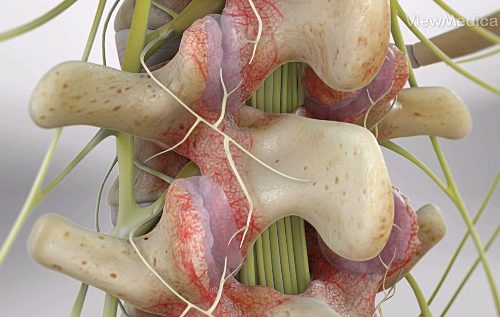
This condition is a deterioration of the facet joints, which help stabilize the spine and limit excessive motion. The facet joints are lined with cartilage and are surrounded by a lubricating capsule that enables the vertebrae to bend and twist.

Facet joint syndrome occurs when the facet joints become stressed and damaged. This damage can occur from everyday wear and tear, injury to the back or neck or because of degeneration of an intervertebral disc.

The cartilage that covers the stressed facet joints gradually wears away. The joints become swollen and stiff. The vertebral bones rub directly against each other, which can lead to the growth of bone spurs along the edges of the facet joints.

Pain from facet joint syndrome differs depending on which region of the spine is damaged. If the cervical, or upper spine is affected, pain may be felt in the neck, shoulders, and upper or middle back. The person may also experience headaches. If the lumbar, or lower spine, is affected pain maybe felt in the lower back, buttocks and back of the thigh.

Facet joint syndrome is first treated conservatively with rest, ice, heat, anti-inflammatory medications, and physical therapy. In addition, facet joint blocks may be administered not only to diagnose facet joint pain but also to treat it. If non-surgical methods fail to relieve pain, a facet rhizotomy or bone fusion may be performed.

The ankles and feet work in unison to enable general mobility and ongoing support to your physical body. A sprain in the foot or ankle is actually an injury that impacts the soft tissue areas. Generally, a sprain happens when a certain injury tears, stretches, or pulls the ligaments that connect the bones together. On the other hand, a fracture is when the bone breaks.

Some of the most common injuries that cause ankle pain are:

  • Bursitis
  • Fractures
  • Sprains
  • Tendonitis

Some diseases can also lead to ankle pain. These include:

  • Arthritis
  • Flatfoot
  • Gout
  • Infections such as cellulitis and osteomyelitis

Steroid injections typically can relieve foot and ankle pain. Your provider uses a needle to inject anti-inflammatory medication directly into your joint. Cortisone shots reduce inflammation and pain.


The vertebrae in your spine are protected by flexible discs that absorb the shocks of your movements. The discs also make it possible for your spine to bend and twist.

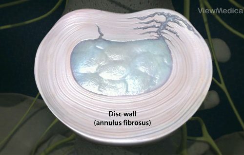
Due to degenerative diseases or the natural changes of aging, discs can wear down and no longer provide adequate protection for the vertebrae. Wear and tear can also force the soft center of the disc through the outer wall, a condition known as a herniated disc.

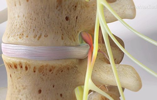
If the protruding center of the disc presses on the surrounding spinal nerves, you can experience chronic pain that worsens over time. The symptoms of a herniated disc depend on the location of the affected disc and how much pressure is being put on the nearby nerves.

In most cases, you can experience persistent pain in the area of the herniated disc, such as the neck or lower back. Pain and weakness can also radiate down into your arms or legs, interfering with normal limb function. When a disc herniation presses on the sciatic nerve, you may have pain or shock-like tingling that radiates down through one side of your buttocks and down your leg.

Initially, you may benefit from muscle relaxants and oral pain medication or steroids to alleviate pain and relieve inflammation surrounding the nerves. Corticosteroid injections are also a minimally invasive option that delivers anesthetic medications to help with the pain and steroids to decrease inflammation, which helps relieve pressure on the nerves.

There are a variety of reasons you can develop ongoing pain in your knee, including:

  • Sprains
  • Arthritis
  • Fractures
  • Infections
  • ACL injuries
  • Inflammation
  • Tendon tears

In some cases, excess pressure on the knee joints from being overweight or repetitive motions from sports can result in chronic knee pain and disability.

What treatments are available for knee pain?
Your custom treatment plan focuses on the cause of pain, with the goal to alleviate pain and associated symptoms so you can regain your mobility. Your provider may recommend injections that contain a local anesthetic and steroids. These medications go directly into the joint to reduce inflammation and provide long-lasting pain relief.

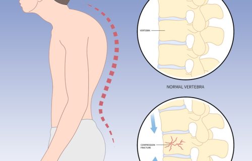
This condition is a deformity of the spine. With it, your vertebrae change from a cylindrical shape to a wedge shape. Your spine may begin to curve forward. Eventually, this gives your upper back a rounded appearance.

Several different causes can result in kyphosis. It may be caused by osteoporosis. This is a loss of bone density. It makes your bones weak and brittle. It can allow your vertebrae to fracture and compress. Kyphosis is also caused by degeneration of your spinal discs. It can be caused by cancer. And it can be caused by birth defects, diseases and syndromes that affect the spine.

If your kyphosis is mild, you may not notice any symptoms. As it progresses, your spine may curve forward. You may experience stiffness. You may have aches in your neck and back. Severe kyphosis may put pressure on your spinal nerves and it may cause your organs to become compressed. This can cause a wide range of problems.

Your neck is part of your spinal column known as the cervical spine. The neck is capable of a wide range of movements and endures carrying the weight of your head.

When you suffer a direct injury to your cervical spine, you can experience ongoing pain and limited mobility in your neck. Degenerative diseases, such as osteoarthritis and spinal stenosis, are also responsible for many cases of chronic neck pain.

In some cases, moving your neck wrong or sleeping in an awkward position can result in a pinched nerve that causes pain and limited range of motion.

If chronic neck pain is the result of a spinal condition, like a herniated disc or spinal stenosis, your provider may recommend cervical epidural steroid injections. This type of treatment delivers anti-inflammatory steroids directly into the epidural space of your cervical spine.

Facet joint injections are also available to address inflamed or irritated joints on both sides of the spine. Injections use a combination of steroids and a local anesthetic to reduce inflammation and block pain signals from traveling to the brain.


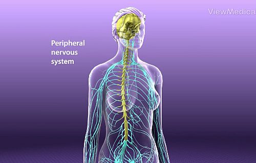
This condition is a problem with the peripheral nervous system. These are the nerves that branch out from your brain and spinal cord and travel to all of the other parts of your body.

This condition results from damage to your peripheral nerves. These nerves can be damaged by diabetes. They can be harmed by disorders of the kidneys, liver and thyroid. They can be damaged by infections and by traumatic injury. Alcoholism, toxins, vitamin deficiencies and other factors can also harm these nerves.

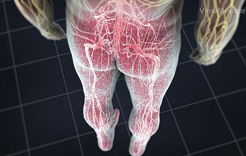
Because the peripheral nerves play a role in so many functions in your body, peripheral neuralgia can cause a wide range of problems. You may have numbness, tingling and painful sensations in your extremities. You may have problems with strength and coordination. You may have cramps and spasms in your muscles. You may be sensitive to heat and touch. You may experience problems with your digestive and urinary systems. Your heart rate and blood pressure may be affected.

This condition, also called “failed back syndrome”, is a type of chronic pain. It can develop in some people after spine surgery.

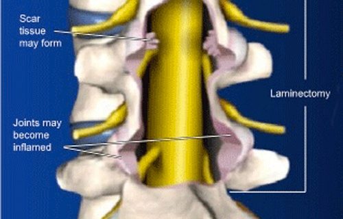
This pain most often develops after a laminectomy procedure. This is the removal of bone at the rear of your vertebrae. The procedure is done to relieve pressure on your spinal nerves. But after a laminectomy, bone or soft tissue may still press on these nerves. Scar tissue may form, and spinal joints may be irritated and inflamed. Pain from any of these issues may be called “post-laminectomy syndrome”.

Symptoms may include pain in your back at the site of your surgery. The pain may also radiate down to your buttock and leg. This pain may feel sharp, or it may feel dull and achy.

Persistent postsurgical pain may be one of the most common problems no one is talking about. It’s estimated that 10-50% of all surgery patients develop chronic postoperative pain that continues long after their body heals.

It’s impossible to predict the extent of the pain you may feel after surgery because each person has a different level of tolerance. The severity of your pain also depends on the type of surgery. Conventional open surgery with one large incision causes more trauma to your body. By comparison, minimally invasive surgery uses small incisions that may not need to cut through the muscle, and it also causes minimal bleeding.

When your postsurgical pain lasts 2-6 months after your procedure, and all other causes for the pain have been ruled out, you have chronic or persistent postoperative pain (PPOP). Damage to major nerves during surgery is one important cause of PPOP. However, there are many cellular changes that occur in your body during surgery that can contribute to PPOP.

Another key factor that contributes to PPOP is a condition called pain sensitization. When your body sustains the trauma of surgery, plus postsurgical inflammation and pain, then biochemical changes can occur that make your nerves more sensitive to pain.


Sciatica pain is often referred to as a “pinched nerve” and can manifest as a sharp “shock” or as a dull aching or burning sensation. Its severity ranges from mild to debilitating. Even though sciatica originates from the lower back, pain may occur anywhere the sciatic nerve runs through, from the lower buttock along the back of the leg and down to the sole of the foot. In most cases, sciatica is limited to only one side.

Is pain the only symptom of sciatica?
While pain is the major concern when dealing with sciatica, problems with the sciatic nerve can also manifest with other signs and symptoms. Patients with sciatica may feel a variety of other sensations in the leg, including:

  • Weakness
  • Numbness
  • Tingling
  • Abnormal reflexes

These, in turn, may lead to clumsiness and difficulty walking. Bowel and/or urinary changes, such as incontinence, may accompany more serious forms of sciatica.

Unfortunately, in many cases of sciatica, the cause can’t be determined. But it can be treated.


This condition is an abnormal curvature of the spine. It most often develops in early childhood, just before a child reaches puberty.

In most cases, the direct cause of scoliosis is not understood. Genetics may play a role as it can run in families. It can also result from neuromuscular conditions, birth defects and injuries of the spine.

A spine with scoliosis, when viewed from the rear, curves sideways. It may curve into a “C” or an “S” shape. Scoliosis tends to develop gradually, and usually it is not painful. A child may not realize it is happening. A parent may overlook it. As the curvature progresses and becomes more severe, scoliosis may cause changes in the body. The shoulders and hips may become uneven. One shoulder blade may stick out prominently. In very severe cases, the spine may begin to twist, causing back pain. The ribcage may appear uneven. This can limit the lungs’ ability to expand, affecting breathing.

Most people who have scoliosis do not need treatment. When it is detected, it can be monitored closely to make sure it isn’t getting worse. A doctor may recommend an orthopedic brace to prevent further curvature.

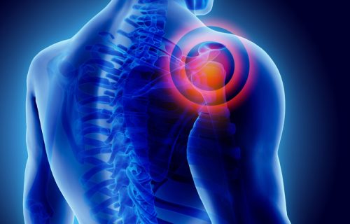
Your shoulders are the joints where your upper-arm bones (humerus), shoulder blades (scapula), and collarbone (clavicle) meet. The humerus fits into the rounded socket of the scapula on each side of your body. Each shoulder is held in place by a group of four muscles and tendons, called a rotator cuff, which covers and protects the humerus and lets you raise and move your arm.

Common shoulder injuries include:

  • Dislocation
  • Separation
  • Fracture
  • Cartilage tear
  • Rotate cuff tear
  • Frozen shoulder
  • Impingement
  • Bursitis


Spinal stenosis is a narrowing of the spine, often resulting from degenerative diseases, such as osteoarthritis. As the spinal vertebrae and their protective discs wear down, they can put pressure on the spinal cord and nerves. Pressure on the nerves can also result as the ligaments in your back thicken as you age, or if you have herniated discs due to injury or disease.

This pressure causes pain and a variety of symptoms that interfere with your mobility and overall quality of life. Your risk for spinal stenosis increases with age or if you suffer trauma to the spine from a fall or auto accident.

Some people can have spinal stenosis without any symptoms, but typically symptoms of the condition develop gradually and worsen over time.

As a result of spinal stenosis, you may experience persistent pain in your back or neck, depending on where the spinal narrowing occurs. Pain can also radiate into your arms and legs, which can interfere with your daily activities and your mobility.

Pain Institute of Southern Arizona continues to be on the cutting edge of medicine, offering a ground breaking treatment for Lumbar Spinal Stenosis. The procedure is called Vertiflex and it is a long-lasting solution to the symptoms of lower back and leg pain.

The procedure is a simple, minimally invasive treatment which can provide effective long-term relief by lifting pressure off the nerves to alleviate leg and back pain:

  • FDA approved
  • Small incision
  • Out-patient procedure
  • Does not require removal of bone or tissue
  • Speedy recovery time
  • Low risk of infection or complications
  • More than 10,000 patients nationwide treated since 2016

The Vertiflex procedure uses a small spacer designed to relieve pressure on the nerves. Your physician will place the spacer between the spinous processes through a small tube that is approximately the size of a dime. Once the spacer is placed, it is intended to relieve pressure off the nerves and reduce the pain, without having to lean forward or sit down.


This condition occurs when a lumbar vertebra slips out of place. It slides forward, distorting the shape of your spine. This may compress the nerves in the spinal canal. The nerves that exit the foramen (open spaces on the sides of your vertebrae) may also be compressed. These compressed nerves can cause pain and other problems.

Spondylolisthesis has a variety of causes. In children, it is often due to a birth defect in that area of the spine. Some people develop this condition because of an overuse injury called “spondylolysis.” This is a stress fracture of the vertebral bone. In adults, arthritis and the loss of disc elasticity that results from aging are the most common causes of spondylolisthesis.

Less commonly, spondylolisthesis can result from a sudden injury that leads to a broken vertebra. Diseases or tumors that weaken the spine can also result in spondylolisthesis.

Symptoms vary from person to person. Many people who have this condition have no symptoms at all. If you do have symptoms, you may experience pain in your lower back. You may have hamstring spasms. Pain may spread down your leg to your foot. You may also have foot numbness and tingling.

Pain Treatments

Click on each treatment to open


This is an outpatient procedure where the physician numbs the arm and hand with an injection of local anesthetic. Typically, it is used before or after surgery on the elbow, forearm or hand.

The patient lies down to expose the armpit. The area is cleaned and sterilized, and a local anesthetic is administered to numb the tissue at the injection site. The physician uses ultrasound or an electrical nerve stimulator to locate the brachial plexus nerves and the axillary artery, which pass through the upper arm. A needle is carefully guided through the armpit and down to the nerves. Anesthetic is slowly injected through the needle, bathing the nerves and temporarily blocking sensation in the arm.


This outpatient procedure is an injection of asteroid-anesthetic medication through an opening in the sacrum. The medication can reduce swelling and inflammation of irritated spinal nerves. The injection takes only a few minutes to complete.

The physician administers a local anesthetic to numb the skin and the tissue above the small opening at the base of the sacrum. This opening is called the sacral hiatus. When the area is numb, the physician guides a needle through the sacral hiatus and into the caudal epidural space. This is the open space in the sacrum where the irritated nerve roots are located.

The physician injects contrast solution through the needle. The physician uses a fluoroscope (a type of x-ray device) to confirm that the tip of the needle is positioned correctly within the epidural space. After the needle’s position has been confirmed, the physician injects a steroid-anesthetic medication. This medication bathes the irritated nerve roots and alleviate pain.


This procedure temporarily disrupts the nerves of the celiac plexus. These nerves branch away from your spine and connect to the organs in your abdomen. Pain signals caused by conditions such as pancreatitis or pancreatic cancer travel through these nerves on the way to your brain. A celiac plexus block can relieve your pain.

In preparation for the block, you are positioned face down on a table. You are given medication to make you feel relaxed. The skin on your back is numbed and the physician inserts a needle through your back and carefully pushes it to the celiac plexus nerves. The needle may be guided with and x-ray device called a fluoroscope. This provides video images. The physician may inject contrast dye to make sure the needle is placed correctly.

The physician injects anesthetic to numb your nerves. If your block is being performed as a test, this may be the end of your procedure. But if your block is being used to treat your pain for an extended period, the physician may inject a substance to disrupt these nerves. This may be repeated to treat the nerves on the other side of your spine.

This injection relieves pain caused by a pinched nerve (or nerves). Conditions such as herniated discs and spinal stenosis can compress nerves, causing inflammation and pain. The medication injected helps decrease the swelling of nerves.

Using x-ray guidance, also called fluoroscopy, the physician guides a needle to the painful area. Contrast dye is injected into the space to make sure the needle is properly positioned near the irritated nerve or nerves.

A combination of an anesthetic and cortisone steroid solution is injected. The steroid is an anti-inflammatory medication. It is absorbed by the inflamed nerves to decrease swelling and relieve pressure. The needle is removed and a small bandage is applied.

In this outpatient procedure, the patient sits or lies down to expose the back. The back is cleaned and sterilized, and a local anesthetic is administered to numb the tissue of the injection site down to the spinal column. The physician uses an x-ray device called a fluoroscope to guide a needle down to the vertebra that is causing the pain. The needle is carefully pushed into the epidural space, which is the area surrounding the spinal cord. A contrast solution is injected to confirm the needle’s position.

The physician injects a steroid-anesthetic mix into the epidural space, bathing the painful areas with soothing medication. The mixture will help reduce inflammation and reduce pain. The needle is removed, and the injection site is covered with a small bandage. Extended pain relief usually begins within three to five days of the injection. In some cases, it may be necessary to repeat the procedure as many as three times to get the full benefit of the medication. However, many patients get significant relief from only one or two injections.


The facet joints, found on both sides of the back of the spine, can become painfully irritated or inflamed. A facet joint injection may help diagnose the source of a patient’s pain. It can also relieve pain and inflammation.

In preparation for the procedure, the physician numbs the skin and tissue above the facet joint with an injection of local anesthetic. With the aid of an x-ray device called a fluoroscope, the physician guides a needle through the numbed tissue and into the facet joint. Contrast dye is injected into the joint to confirm the needle’s placement. Once the needle is positioned properly, the physician injects a soothing mixture of numbing anesthetic and anti-inflammatory steroid medication. One or more facet joints may be treated. If this causes the pain to subside, it suggests that the facet joint (or joints) injected were the cause of pain.

Back or neck pain may disappear immediately after a successful injection because of the anesthetic that is administered. As this anesthetic wears off, pain may return. The steroid will begin to take effect in the days after the injection. The steroid will reduce inflammation and pain.

The Intracept Procedure is a minimally invasive, implant free procedure that preserves the overall structure of the spine. The Intracept Procedure is a same-day, outpatient procedure. Patients are under anesthesia, and the procedure generally lasts an hour. The procedure is FDA-cleared and is proven in multiple studies to be safe, effective, and durable.

Learn more.

Joint injection procedures are performed to increase mobility and relieve joint bone and joint pain due to injury, arthritis or other causes.

In this outpatient procedure, you lie on your back and your skin is numbed. The doctor pushes a very small needle through the numbed skin and down to your hip joint. Ultrasound can be used to guide the needle or, a video x-ray device called a fluoroscope can be used. If so, the doctor will inject some contrast dye to help make sure the needle is in your joint. The soothing medicine is injected and it bathes your joint.


This minimally-invasive procedure repairs a vertebral compression fracture and helps restore the spine’s natural shape. Some patients experience rapid pain relief after the procedure.

Before the procedure, you are sedated. The physician guides a needle through the skin of your back and into your fractured vertebra. A special x-ray device called a fluoroscope helps the physician position the needle.

A balloon device is placed through the needle and into the vertebral body. The physician carefully inflates this balloon to expand the fractured bone. When the balloon is deflated, it leaves a cavity in the middle of the vertebral body. The balloon is then removed. The physician then injects bone cement through the needle, which fills the cavity. The cement hardens inside the vertebral body, stabilizing the fracture. When the procedure is complete, the needle is removed. The opening in your skin is closed.


This procedure is an injection that numbs branches of nerves in your lower back. It helps doctors find and treat a number of problems linked to these nerves. Usually, a series of injections is needed to treat a problem.

The sympathetic nerves travel along both sides of your spine. They are associated with a wide range of functions that you don’t consciously control. These include your circulation, digestion and sweat production.

In preparation for the procedure, you lie on your stomach or your side. You are given medicine to make you feel relaxed. The skin and tissue at the injection site is numbed. The physician inserts a needle and carefully guides it to the sympathetic nerves. The physician typically uses an x-ray device called a fluoroscope which shows a video image of the needle’s position. Contrast dye may be injected to help confirm that the needle is placed correctly.

Next, the physician injects soothing medicine which bathes the nerves. It can numb the nerves and reduce inflammation. If these nerves have been a source of pain, the medicine can relieve it. When the procedure is complete, the needle is removed and the injection site is covered with a bandage.

Medication management is a common method of pain treatment. Medications can help patients in pain lead a more active life. With proper consultation and diagnosis, a medication management plan can be put into place and monitored on a regular basis.


This minimally invasive procedure restricts pain signals to the brain and helps combat the debilitating symptoms of conditions such as arthritic joint pain, neck pain, and lower back pain.

During the procedure, radiofrequency needles are placed alongside the nerve that goes to the joint. The physician makes the joint numb with local anesthetic and then heats the needles with radiofrequency waves to burn the nerves that go to the joints. This process damages the nerves, restricting their ability to send pain signals to the brain.

Patients are typically sore for a couple days after the procedure and it takes two to six weeks for the procedure to become effective. Patients typically experience 80% to 100% pain relief that can last for six months to several years. Results of radiofrequency ablation are not permanent because it only temporarily interferes with how the spinal column sends pain signals to the brain. If the patient experiences sufficient relief but the pain returns, the procedure can be repeated.


When your chronic pain fails to respond to conventional medical care, you may find relief from one of today’s most innovative technologies called a spinal cord stimulator. Spinal cord stimulators use mild electrical impulses to block or mask the pain signals transmitted by nerves. When any part of your body is in pain, sensory nerves pick up the message and transmit it to your brain. You only feel the pain after your brain receives and interprets the message.

Spinal cord stimulators effectively reduce your pain by stopping the message from reaching your brain or by masking it so your brain doesn’t understand the message. The source of your pain is still there, but your pain is significantly diminished.

Spinal cord stimulators consist of three parts: a small generator, a wire with multiple electrodes, and a controller. The generator, which produces the electrical impulses, is implanted under the skin of your abdomen, upper buttocks, or chest. The long, flexible wire is threaded along your spine. Your physician uses fluoroscopic imaging to see the wire and place its electrodes near the spinal nerves responsible for sending the pain signals to your brain. Your physician uses the remote controller to initially program the strength and frequency of the electrical impulses. Then you’ll use it to turn your spinal cord stimulator on and off.

You’ll have a trial period with the spinal cord stimulator to determine if it works for you. During your trial, your physician implants the wires, but you’ll wear the generator around your waist. After about one week of wearing the stimulator, you’ll know if it’s effectively relieving your pain. At that point, you can decide to have the generator implanted and keep the device, or your physician can easily remove the wires.


A stellate ganglion nerve block is an injection that numbs branches of nerves in your neck. This helps doctors find and treat a number of problems linked to the nerves. Treatment may require a series of injections.

The stellate ganglion nerves travel along both sides of your spine. They are associated with a wide range of bodily functions that you don’t consciously control. These include blushing, heart rate, sweating and the dilation of your pupils.

In preparation for the procedure, you lie on your back. You are given medicine to make you feel relaxed. The skin and tissue of your neck is numbed. The physician inserts a needle and carefully guides it to the nerves of the stellate ganglion. The physician typically uses an x-ray device called a fluoroscope which shows a video image of the needle’s position. Contrast dye may be injected to help confirm that the needle is placed correctly. Next, the physician injects soothing medicine which bathes the nerves. It can numb the nerves and reduce inflammation. If these nerves have been a source of pain, the medicine can relieve it.


A trigger point is a tight knot that develops in a muscle or the fascia, which is the connective tissues covering your muscles and organs. You can develop a single trigger point, or several knots, along a band of muscles. If your trigger point causes chronic pain, the condition is called myofascial pain syndrome.

There are two types of trigger points:

Active trigger points
Active trigger points are very sensitive and painful, often causing pain even when you’re resting. You may also develop muscle weakness, limited movement, and referred pain, which is pain that’s felt elsewhere in your body when the trigger point is pressed. Pressure on the trigger point may also cause an automatic muscle twitch.

Latent trigger points
Latent trigger points may hurt when you press on the knotted area, but they’re less painful than active trigger points.

Trigger points are usually caused by:

  • Repetitive muscle movements
  • Lifting heavy weights or items
  • Direct injury
  • Poor posture
  • Tense muscles

Although trigger points most often develop due to overusing or placing excessive stress on your muscles, they can also occur if you stay inactive for too long.

A trigger point injection contains a local anesthetic. The local anesthetic blocks pain signals, providing immediate relief. Steroids alleviate pain by reducing inflammation. It takes several days to feel the effect of steroids, but they provide longer-lasting relief.

During a trigger point injection, your doctor finds the trigger point, and may apply a topical anesthetic on the skin above the knotted muscle. Then the needle is inserted into the trigger point and the medication is injected. If the trigger point doesn’t fully relax after one injection, your doctor may slightly reposition the needle and deliver another injection.

The Vertiflex procedure is a ground-breaking treatment for Lumbar Spinal Stenosis. It is long-lasting solution to they symptoms of lower back and leg pain.

The procedure is a simple, minimally invasive treatment which can provide effective long-term relief by lifting pressure off the nerves to alleviate leg and back pain:

  • FDA approved
  • Small incision
  • Out-patient procedure
  • Does not require removal of bone or tissue
  • Speedy recovery time
  • Low risk of infection or complications

The Vertiflex procedure uses a small spacer designed to relieve pressure on the nerves. Your physician will place the spacer between the spinous processes through a small tube that is approximately the size of a dime. Once the spacer is placed, it is intended to relieve pressure off the nerves, and reduce the pain, without having to lean forward or sit down.

Schedule Your Appointment Today!

Call 520-999-9000 or click Request Appointment Online button

Translate